| Provider | |
|---|---|
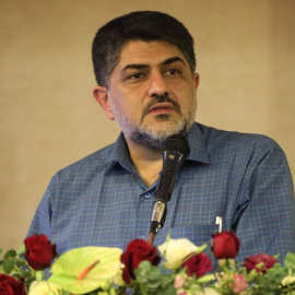
Hossein Rabbani , PhDFull Professor
School :School of Advanced Technologies in MedicineDepartment :Department of Bioelectrics and Biomedical EngineeringResearch Center :Medical Image & Signal Processingwork address :https://goo.gl/maps/WxjBDo4MyhyEducation :2013 –2014 Postdoctoral Research Fellow, Duke Eye Center, Durham, United States. 2011 Postdoctoral Research Scholar, The University of Iowa, Iowa, United States. 2007 Visiting Research Scholar, Electrical & Computer Engineering Department, Queen’s University, Kingston, Ontario, Canada. 2002 – 2008 Doctoral Researches, Bioelectrics, Department of Biomedical Engineering, Amirkabir University of Technology (Tehran Polytechnic), Tehran, Iran. 2000 – 2002 Master of Science, Bioelectrics, Department of Biomedical Engineering, Amirkabir University of Technology (Tehran Polytechnic), Tehran, Iran. 1997 – 2000 Bachelor of Science, Communications (1st rank), Department of Electrical and Computer Engineering, Isfahan University of Technology, Isfahan, Iran. Relevant Work Experience :Hossein Rabbani (Senior Member, IEEE) received the B.Sc. degree (Hons.) in electrical engineering (communications) from Isfahan University of Technology, Isfahan, Iran, in 2000, and the M.Sc. and Ph.D. degrees in bioelectrical engineering from the Amirkabir University of Technology (Tehran Polytechnic), Tehran, Iran, in 2002 and 2008, respectively. In 2007, he was with the Department of Electrical and Computer Engineering, Queen’s University, Kingston, ON, Canada, as a Visiting Researcher. In 2011, he was with the University of Iowa, IA, USA, as a Postdoctoral Research Scholar. From 2013 to 2014, he was with Duke University Eye Center as a Postdoctoral Fellow and from 2020 to 2023, he was with the University of Göttingen, Lower Saxony, Germany (the winner of Georg Forster Fellowship for Experienced Researchers by Alexander von Humboldt Foundation). He is currently a Professor with the Biomedical Engineering Department and the Medical Image and Signal Processing Research Center, Isfahan University of Medical Sciences, Isfahan, Iran. His main research interests include medical image analysis and modeling, statistical (multidimensional) signal processing, sparse transforms, and image restoration, which more than 200 papers and book chapters have been published by him as the author or the co-author in these areas. He is the Editor-in-Chief of Journal of Medical Signals and Sensors (JMSS) and Associate Editor of IEEE Transactions on Medical Imaging. For more information please visit: • https://hrabbani.site123.me • http://misp.mui.ac.ir • https://sites.google.com/site/hosseinrabbanikhorasgani Professional Memberships & Qualification :B.Sc. Degree with the honor of the 1st rank from Isfahan University of Technology. M.Sc. Degree with the honor of the 3rd rank from Amirkabir University of Technology. 1st rank in the Ph.D. entrance exam of Amirkabir University of Technology in bioelectrics. 1st rank in the Ph.D. entrance exam of Isfahan University of Technology in communication engineering. Outstanding selected Ph.D. thesis by Iranian Society for Biomedical Engineering. The winner of national prize of “Young Assistant Professors”. IEEE, Elected Senior Member, 2013. The Winner of Avicenna Research Award, Isfahan Univ. of Med. Sciences (2019). The recipient of Georg Forster research fellowship for experienced researchers, Alexander von Humboldt Foundation (2019). The winner of Seed Money Grant with Switzerland Leading House. Extra Curricular Activities/ Interest(personal url) :Grants & Awards :Seed Money Grant (Swiss): A feasibility study to develop an OCT-based ocular health kiosk to diagnose Diabetic Retinopathy (20,000 CHF) NIMAD: Alignment of Optic Nerve Head OCT B-Scans in Right and Left Eyes for Glaucoma Detection (262,500,000 Rials) National Elites Foundation: OCT and Angiography Image Registration of Diabetic Retinopathy Patients (600,000,000 Rials) INSF: Intelligent System for Abnormality Detection and Intra-Retinal Layer Segmentation of Optical Coherence Tomography Images Using 3D Convolutional Neural Networks (324,000,000 Rials) Isfahan University of Medical Sciences: Morphological Component Analysis of Optical Coherence Tomography (OCT) data (150,000,000 Rials) National Elites Foundation: 3D intra-retinal layer segmentation in OCT data using modified live wire (600,000,000 Rials) National Elites Foundation: Synchronized analysis of EEG, MRI images, and SPECT images of patients suffering from seizure (600,000,000 Rials) Microelectronics Council: Hardware Implementation of Medical Image Processing Algorithms (720,000,000 Rials) National Elites Foundation: Medical Image Modeling by Spherically Invariant Random Variables (200,000,000 Rials) Patents :Presentations & Poster Sessions :· Combination of graph based algorithms and time-frequency methods for processing of OCTs · Seeking an appropriate feature extraction method for breast cancer recurrence prediction based on microarray gene expression data · A new model based on Gaussianization of OCT data · Automatic analysis of features of AMD in OCT images using 3D curvelet transform · Automatic detection of acute myeloid leukemia in microscopic images using dictionary learning · Automatic diagnosis of Mild Cognitive Impairment (MCI) by dictionary learning -based analysis of EEG signals · 3D OCT Classification by Deep Learning · Fully automated segmentation of fluid/cyst regions in OCT images using neutrosophic sets and graph algorithms · Polyp detection/segmentation in video colonoscopy by convolutional neural network · Image restoration using Gaussian mixture models with neighborhood nonlocal clustering · Evaluation of the symmetricity of cup to disk ratio in left and right eyes of normal subjects · Mosaicing macula OCT images and OCT optical disk · Designing a dictionary for OCT images based on K-SVD algorithm using texture characteristics of retinal layers for image segmentation · Automatic segmentation of corneal layer boundaries in OCT images and obtaining 3D maps of the entire thickness of cornea and inner layers · Automatic detection of leishman bodies in bone marrow samples from patients with visceral leishmaniasis using level set method · Evaluation of asymmetricity of right and left eyes of normal subjects using extracted features from optical coherence tomography (OCT) and color fundus images · Automatic diagnosis of malaria based on complete circle-ellipse fitting search algorithm · Automatic segmentation and recognition of lung nodules in thoracic CT images using active contour modeling and convex hull · Segmentation of enhanced depth imaging optical coherence tomography (EDI-OCT) images using graph cut algorithm based on Gaussian mixture model of wavelet features · Forming projection images from retinal layers on the 3D OCT data and fusion of them using curvelet transform to form an optimal projection image · Evaluation of image pre-compensation methods for enhancing visual efficiency in the presence of higher order ocular optical aberrations · Extraction of 15-lead ECG signal from vectorcardiogram (VCG) signal using partial linear transformation for providing information from posterior side of the heart · Detection of foveal avascular zone (FAZ) based on curvelet transform for grading of diabetic retinopathy · Extraction of nucleolus candidate zone in white blood cells of peripheral blood smear images using curvelet transform · A comparison between hp version of finite element method with EIDORS for electrical impedance tomography · A comparison between ECG and VCG signals for detection of ischemia location · Estimation of somatosensory evoked potentials with multiadaptive filters · A contourlet-based watermarking method for medical images · Automatic detection of diabetic retinopathy by extraction of retinal image features in curvelet domain · Estimating depth of anesthesia based on wavelet transform and neuro-fuzzy systems · Microcalcification detection in mammographic images using fractal model in wavelet domain · Persian script character recognition using PCA · A comparison between ECG and VCG for detection of ischemia · Automatic detection and recognition of lung nodule in CT image based on active contour · Complexity analysis of EEG signals for Mild Cognitive Impairment (MCI) diagnosis · 3D segmentation of proximal enamel lesions in micro-ct images · Automatic analysis of tracheal acoustic signals for apnea detection and introducing new clinical indices of depth of sedation · Statistical modeling of 3D OCT data by mixture model · Transform based ellipse detection in microscopic images using elliptical basis functions · Automatic extraction and recognition of myeloma cell in microscopic bone marrow aspiration images · Extraction of candida fungus from pap smear images based on ridgelet transform for vulvovaginal candidiasis diagnosis · Extraction of vessels, optic disc and fovea avascular zone from fundus fluorescein angiogram based on Hessian analysis of directional curvelet subbands · Medical image compression with multi-wavelet · A new adaptive technique for fast and accurate estimation of SSAEP Research Experience :1. M. Mokhtari, H. Rabbani*, A. Mehridehnavi, R. Kafieh, M. Akhlaghi, M. Pourazizi, L. Fang, "Local comparison of cup to disc ratio in right and left eyes based on fusion of color fundus images and OCT B-scans", Information Fusion, vol. 51, pp. 30-41, 2019. 2. M. Lashgari, M. Shahmoradi, H. Rabbani*, M. Swain, "Missing Surface Estimation Based on Modified Tikhonov Regularization: Application for Destructed Dental Tissue," IEEE Transactions on Image Processing, vol. 27, no.5, pp. 2433-2446, 2018. 3. R. Rasti, H. Rabbani*, A. Mehridehnavi and F. Hajizadeh, "Macular OCT Classification using a Multi-Scale Convolutional Neural Network Ensemble," IEEE Transactions on Medical Imaging, vol. 37, no. 4, pp. 1024-1034, 2018. 4. A. Rashno, D. D. Koozekanani, P. M. Drayna, B. Nazari, S. Sadri, H. Rabbani, K. K. Parhi*, "Fully-Automated Segmentation of Fluid/Cyst Regions in Optical Coherence Tomography Images with Diabetic Macular Edema using Neutrosophic Sets and Graph Algorithms," IEEE Transactions on Biomedical Engineering, vol. 65, no. 5, pp. 989-1001, May 2018. 5. M.J. Allingham*, D. Mukherjee, E.B. Lally, H. Rabbani, P.S. Mettu, S.W. Cousins, S. Farsiu, "A Quantitative Approach to Predict Differential Effects of Anti-VEGF Treatment on Diffuse and Focal Leakage in Patients with Diabetic Macular Edema - A Pilot Study", Translational Vision Science & Technology, vol. 7, no. 6, 2017. 6. Z. Amini, H. Rabbani*, "Statistical Modeling of Retinal Optical Coherence Tomography", IEEE Transactions on Medical Imaging, vol. 35, no. 6, pp. 1544-1554, 2016. 7. O. Sarrafzadeh, H. Rabbani, A. Mehri Dehnavi*, A. Talebi, "Analyzing Features by SWLDA for the Cassification of HEp-2 Cell Images Using GMM", Pattern Recognition Letters, vol. 82, part 1, pp. 44-85, Oct. 2016. 8. M. Shahmoradi, M. Lashgari, H. Rabbani*, J. Qin, M. Swain, "A comparative study of new and current methods for dental micro-CT image denoising", Dentomaxillofacial Radiology, vol. 45, no. 3, 2016. 9. M. Niknejad, H. Rabbani*, M. Babaei-Zadeh, "Image Restoration Using Gaussian Mixture Models with Spatially Constrained Patch Clustering," IEEE Transactions on Image Processing, vol. 24, no. 11, pp. 3624-3636, Nov. 2015. 10. R. Kafieh, H. Rabbani*, I. Selesnick, “Three Dimensional Data-Driven Multi Scale Atomic Representation of Optical Coherence Tomography”, IEEE Transactions on Medical Imaging, vol. 34, no. 5, pp. 1042-1062, 2015. 11. H. Rabbani*, M.J. Allingham, P.S. Mettu, S.W. Cousins, S. Farsiu, “Fully Automatic Segmentation of Fluorescein Leakage in Subjects with Diabetic Macular Edema”, Investigative Ophthalmology and Visual Science, vol. 56, no. 3, pp. 1482-1492, 2015. 12. MR. Sehhati*, A. Mehri, H. Rabbani, M. Pourhossein, “Stable Gene Signature Selection for Prediction of Breast Cancer Recurrence Using Joint Mutual Information”, IEEE/ACM Transactions on Computational Biology and Bioinformatics, vol. 12, no. 6, pp. 1440-1448, 2015. 13. R. Kafieh, H. Rabbani*, M. Sonka, M. D. Abramoff, “Intra-Retinal Layer Segmentation of 3D Optical Coherence Tomography Using Coarse Grained Diffusion Map”, Medical Image Analysis, vol. 17, no. 8, pp. 907-928, Dec. 2013. 14. R. Kafieh, H. Rabbani*, S. Kermani, "A Review of Algorithms for Segmentation of Optical Coherence Tomography from Retina", Journal of Medical Signals and Sensors, vol. 3, no. 1, pp. 45-60, 2013. 15. R. Kafieh, H. Rabbani*, F. Hajizadeh, M. Ommani, S. Kermani, “An Accurate Multimodal 3D Vessel Segmentation Method Based on Brightness Variations on OCT Layers and Curvelet Domain Fundus Image Analysis”, IEEE Transactions on Biomedical Engineering, vol. 60, no. 10, pp. 2815-2823, Oct. 2013. 16. M. Sheikhhosseini, H. Rabbani*, M. Zekri, A. Talebi, “Automatic Diagnosis of Malaria Based on Complete Circle-Ellipse Fitting Search Algorithm”, Journal of Microscopy, vol. 252, no. 3, pp. 189-203, Dec. 2013. 17. M. Esmaeili, H. Rabbani*, A. Mehri, “Automatic Optic Disk Boundary Extraction by the Use of Curvelet Transform and Deformable Variational Level Set Model”, Pattern Recognition, vol. 47, no. 7, pp. 2832-2842, July 2012. 18. F. Rahimi and H. Rabbani*, “A dual adaptive watermarking scheme in contourlet domain for DICOM images”, BioMedical Engineering OnLine, 10:53, 2011. 19. H. Rabbani, S. Gazor*, R. Nezafat, “Wavelet-Based 3D Medical Image Denoising Using Bivariate Laplacian Mixture Model”, IEEE Transactions on Biomedical Engineering, vol. 56, no. 12, pp.2826-2837, Dec. 2009. 20. H. Rabbani*, “Image Denoising in Steerable Pyramid Domain Based on a Local Laplace Prior”, Pattern Recognition, vol. 42, pp. 2181-2193, Sept. 2009. 21. H. Rabbani, M. Vafadoost, S. Gazor*, P. Abolmaesumi, “Speckle Noise Reduction of Medical Ultrasound Images in Complex Wavelet Domain Using Mixture Priors”, IEEE Transactions on Biomedical Engineering, vol. 55, no.9, pp. 2152-2160, Sept. 2008. Research Interests :· Intra-retinal layer and fluid segmentation of 3D OCT images by deep learning · Classification of ocular images using optimum basis functions (DL & MCA) · OCT modeling: statistical modeling vs. geometrical modeling vs. energy-based modeling · Synchronized analysis of EEG, MRI images, and SPECT images of patients suffering from seizure · 3D Sparse Reconstruction of Cone-beam CT · Multivariate Statistical Modeling of OCT Images · Sparse Representation of PH Monitoring Signals · Energy-based Modeling of OCT Images · Sparse Representation of OCT Images Teaching Experience :Teaching Interests : |

